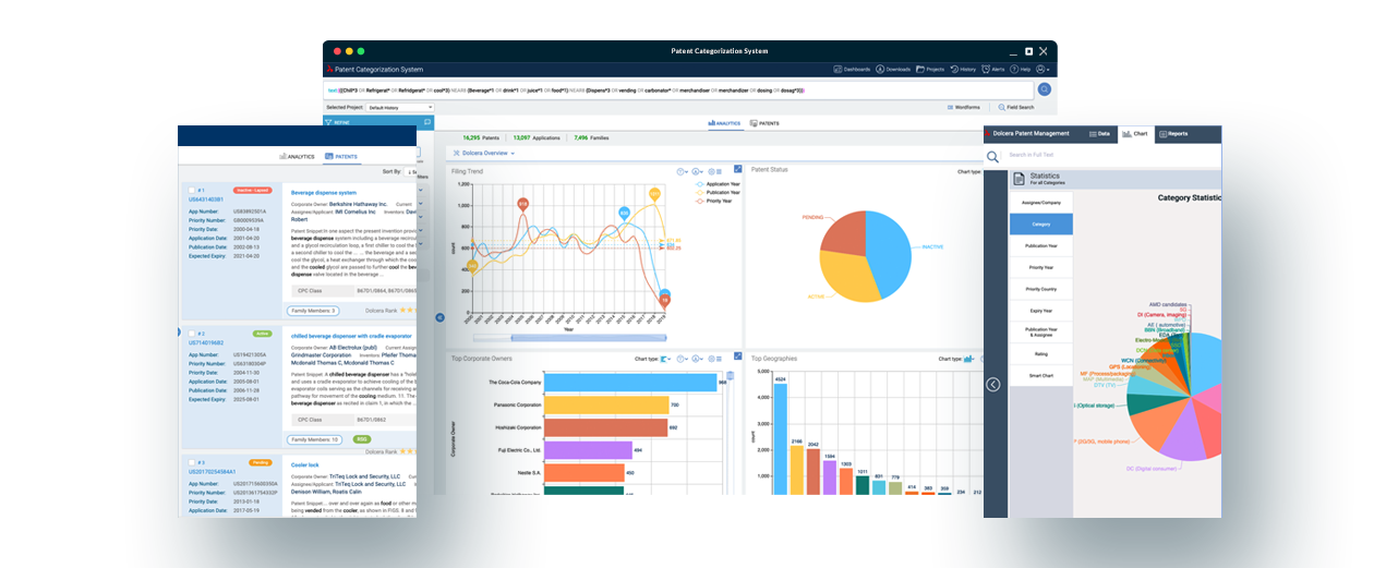
Need an Organ?? Print it!

What’s Organ printing? What’s Bioprinting? Does organ printing really mean what it says in itself?? Pr-in-ting organs?  What do these technologies have to offer? What would the implications of these technologies be on the state of the industry, the general public and the entire humanity? Let’s just break the fourth wall here.  Who’s asking??

Well…To clarify, we are asking these questions in the interest of and to interest the general public and if the latter isn’t lost already (we earnestly hope that is the case) please read up…

Creating living tissue in complex geometries is a challenging issue facing the tissue engineering community.  Traditional tissue engineering techniques result in living tissue of simple, often 2D geometries.  By harnessing the capabilities of Solid-Freeform Fabrication (SFF) – also known as Rapid Prototyping (RP) –living tissue of arbitrary 3D shapes can be created directly from computer-aided design (CAD) data.

Bio-printing is a variant of 3D printing and can be defined as computer-aided, automatic, layer-by-layer deposition, transfer, and patterning of biologically relevant materials.  It is also known by other names such as “computer aided tissue engineering” and “biofabrication”.  In simpler words, bioprinting involves printing devices that deposit biological material.

Organ printing is a variant of bio-printing aiming at producing 3D organs. This is among the most promising advances of regenerative medicine. The 3D- Bioprinter was listed among the TIME magazine’s 50 best inventions of 2010. Most of the 3D printers use a modified version of inkjet printers to deposit dots of “bio ink” (cell suspension with 10 to 30 thousand cells per drop) that coalesce to form layers of organ interrupted by layers of biopaper (hydrogel mimicking the microenvironment of tissue) which is water-soluble.

Step-by-Step process of Organ Printing

Fig1:- Step-by-Step process of Organ Printing [1]


Roadmap to Bioprinting

Roadmap to Bioprinting

Fig 2:- Roadmap to Bioprinting [2]

The 3D bioprinters currently in the market are produced by envisionTEC, Organovo, Tengion, Sciperio, Neatco, etc.

The NovoGen MMX 3D Bioprinter, priced at $200,000, has been developed by Organovo, a company in San Diego that specializes in regenerative medicine, and Invetech, engineering and automation firm in Melbourne, Australia. One of the founders of Organovo, Gabor Forgacs from the University of Missouri, Columbia, says the logic behind applying 3D printing for producing biological organs is “Although morphogenetic processes are under strict genetic control, genes do not create shapes and forms: physical mechanisms and processes do.” Organovo announced the production of first fully bioprinted blood vessels in Dec 2010. [3]

Organovo’s only real competitor, Tengion, holds most of the patents and legal rights to exploit the technology developed by the most successful bio printing scientist to date, Dr. Anthony Atala. Dr. Atala’s lab has grown a variety of human parts including blood vessels, heart valves and bladders, all using bio printing. Based on the same 3D print technology that Organovo uses, Tengion’s prototype printer has already produced several bladders which have been successfully transplanted into humans.

The 3D Bioplotter is produced by German company envisionTEC is a German company producing a range of 3D bioprinters such as 3D Bioplotter, E-Dent (Digital Dental printer), etc.  The 3D Bioplotter is priced at $188,000 and currently used in various laboratories to create various tissue scaffolds. [4]

Work of other research groups on Bioprinting:-

Boland and his coworkers from Clemson University have been producing Bioprinters since 2004.

VAXDesign of Sciperio Inc has a pressure operated 3D Bioprinter with four nozzles in the market.

Roland, Fishman, and Neatco collaboratively produce a pressure operated 3D Bioprinter with two nozzles. [1]

Professor Nakamura from the University of Toyama is currently working with Epson to produce 3D Bioprinters.

Sangeeta Bhatia from MIT together with Jennifer West from Rice University bioprinted living 3D liver constructs using stereolithography.

Tsinghua University group in China also printed liver construct using chitosan-collagen hydrogel.

A research group at Cornell University bioprinted living cartilage construct. [6]

The Bioprinting community meets annually at the International Conference on Bioprinting and Biofabrication. The next conference (the sixth) is to be held in Toyama, Japan in October 2011.

Future and Implications:-

Professor Vladimir Mironov, Director of Medical University of South Carolina(MUSC) , Bioprinting Center says that it would probably take an investment of  $1 billion to print living human organ suitable for clinical implantation.

Also, Dr. Atala says in vivo bioprinting i.e.; bioprinting right into a patient on the site of injury is very much feasible.

As Chair of The Department of Surgery of Stanford University and a leading expert in surgical innovation, Krummel recently wrote: “There is no such thing as a science fiction. There is only science eventuality”. [1]

Here’s to a future when Organ transplantation is hassle-free, affordable, automated, and customized.

Sources:-

1.       http://accessscience.com/content/Tissue-and-organ-printing/YB060455

2.       http://organprint.missouri.edu/PDF/HowToPrintOrgan-slides.pdf

3.       http://www.economist.com/node/15543683?story_id=15543683&subjectID=526354&fsrc=nwl

4.       http://www.envisiontec.de/index.php?id=8

5.       http://www.rapidtoday.com/future.html

Written by:

 Ajay S and Jnanasiddhy

[Post to Twitter] Tweet  [Post to Delicious] Delicious  [Post to Digg] Digg  [Post to Facebook] Facebook 

The Wellness Chip takes clinical diagnosis to newer heights

A simple blood test and there you go….your complete health profile!

Or are you worried about a family cancer history?

The benefit of early diagnosis of such a deadly disease is obvious. The answer is the Wellness Chip®.

Researchers at Somalogic, Inc. have developed a diagnostic technology with high sensitivity and specificity. The Wellness Chip, the brainchild of Larry Gold and his colleagues at Somalogic, will bring together diagnostic tests for various diseases in a single, simple blood test. Currently, Somalogic has licensed its technology to Quest Diagnostics for launching a lung cancer blood test before the end of this year.

What is the Wellness Chip?

The underlying principle is nucleic acid-aptamer based proteomic technology. Somalogic’s proprietary reagents called SOMAmers (Slow Off-rate Modified Aptamers) are single strand stretches of modified nucleic acids generated by a highly selective process called SELEX (Systematic Evolution of Ligands by Exponential Enrichment). SOMAmers are capable of binding specifically to a target of interest, e.g., a marker for a particular disease. SOMAmers to various targets are fabricated on a chip at distinct positions and simultaneously detect several proteins in micro-liter scale samples. This data will then be analyzed by bioinformatics tools.

Contrary to conventional antibodies based diagnostic procedures, SOMAmers are more sensitive and specific and can be made rapidly in weeks rather than months as is the case with antibodies. It will also be economically priced -$100’s not $1000’s.

Why blood test?

Proteins present in the blood are immediate indicators of a person’s state of health.  Diseased tissues secrete proteins in the blood and their identification is essential to early and accurate therapeutic and preventative measures. The total number of blood proteins is about 4000 and the Wellness Chip can detect 1100 of these blood proteins. This protein profile will be analyzed to determine the health status of the individual. Currently available methods detect only about 20 to 30 proteins at a time.

From the laboratory to the market-

The SOMAmer technology has already found its clients in the pharma industry. In 2008, Somalogic entered into research collaboration with Otsuka Pharmaceutical Co., Ltd. for the development of diagnostic tools and to further Otsuka’s research in pharmaceuticals. On the other hand, the task of making the Wellness Chip a reality is entrusted with Quest Diagnostics.

Sources:

1.      http://www.somalogic.com/

2.      Bio.IT World.com. Turning blood into gold: The Wellness Chip

3.      http://www.colorado.edu/mcdb/goldlab/Slide%20Decks/8.%20Steve%20Williams%20slides.pdf

4.      U.S. Patent No. 7709192

5.      Gold L, Ayers D, Bertino J, Bock C, Bock A, et al. (2010) Aptamer-Based Multiplexed Proteomic Technology for Biomarker Discovery. PLoS ONE 5(12): e15004

6.      Ostroff RM, Bigbee WL, Franklin W, Gold L, Mehan M, et al. (2010) Unlocking Biomarker Discovery: Large Scale Application of Aptamer Proteomic Technology for Early Detection of Lung Cancer. PLoS ONE 5(12): e15003.


Written by:

Shweta Kumari

[Post to Twitter] Tweet  [Post to Delicious] Delicious  [Post to Digg] Digg  [Post to Facebook] Facebook 

Impotent Warriors- War against Malaria

Mosquitoes are deadly little creatures troubling mankind since time immemorial. These are carriers of a deadly parasite responsible for the spread of Malaria- claiming millions of deaths worldwide. Despite enormous efforts to curb this disease, not much has been achieved and scientific communities are striving to come up with an effective defense mechanism. A new study led by Dr Flaminia Catteruccia from the Department of Life Sciences at Imperial College London, focuses on exploiting the reproductive mechanism of mosquitoes- a new dimension in malaria research.Interestingly, Anopheles gambiae, a species primarily responsible for transmission of malaria in Africa, mates only once in its lifetime. Scientists believe that interfering with mosquito reproduction could be a potential defense against malaria.  “In the fight against malaria, many hope that the ability to genetically control the mosquito vector will one day be a key part of our armory,” said Flaminia Catteruccia.

This could be achieved either by inhibiting the reproduction process or sterilizing the male mosquitoes by genetically modifying them to neutralize a gene required for sperm production.

In 2009 , research led by Flaminia demonstrated that knocking off a key enzyme called Transglutaminase results in an impaired mating plug, a coagulate of seminal fluid and proteins required to seal the male sperm inside female, and thus disrupts the reproductive process thereby providing a potential means to combat malaria.

Dr Catteruccia concludes: “If in the future we can develop an inhibitor that prevents the coagulating enzyme doing its job inside male A. gambiae mosquitoes in such a way that can be deployed easily in the field — for example in the form of a spray as it is done with insecticides — then we could effectively induce sterility in female mosquitoes in the wild. This could provide a new way of limiting the population of this species of mosquito, and could be one more weapon in the arsenal against malaria.”

Recently, a path breaking study (published in PNAS journal) reveals that genetically modified mosquitoes with inability to produce sperms mate successfully with the females as compared to the normal counterpart and crucially, this modification did not interfere with any other sexual function or behavior in either the female or the male, they explained in their study.

“Spermless males behave exactly like those with sperm,” Catteruccia says. “We saw no difference in their ability to compete.” She further added that releasing genetically modified, spermless male mosquitoes into the wild could in future help to prevent malaria transmission and reduce the chances of large outbreaks of the killer disease.

Furthermore, Elena Levashina who studies malaria carrying mosquitoes, a leading scientist at the Institute of Molecular and Cellular Biology in Strasbourg comments that “this is a crucial scientific demonstration and a huge step forward.”

This seemingly promising strategy to deal with outbreak of malaria is undoubtedly a major advancement and a potential weapon against malaria. However, these studies are in their initial stage and have been tested in field trials only and further experiments have to be done to use it on a large scale.

SOURCES:

Imperial College London. “Meddling in mosquitoes’ sex lives could help stop the spread of malaria.” ScienceDaily, 22 Dec. 2009. Web. 12 Aug. 2011.

Imperial College London. “Mosquitoes can’t spot a spermless mate.” ScienceDaily, 10 Aug. 2011. Web. 12 Aug. 2011.

Nature NEWS. “Female mosquitoes tricked by spermless males.” Published online 8 August 2011 | Nature | doi:10.1038/news.2011.467

Journal References:

  1. Thailayil J, Magnusson K, Godfray HCJ, Crisanti A, Catteruccia F. Spermless males elicit large-scale female responses to mating in the malaria mosquito Anopheles gambiae. PNAS, 2011.
  1. Rogers DW, Baldini F, Battaglia F, Panico M, Dell A, Morris HR, Catteruccia F. Transglutaminase-mediated semen coagulation controls sperm storage in the malaria mosquito. PLoS Biol. 2009.

Written by:

Abu Rafay

[Post to Twitter] Tweet  [Post to Delicious] Delicious  [Post to Digg] Digg  [Post to Facebook] Facebook 

Tweet links powered by Tweet This v1.4.1, a WordPress plugin for Twitter.